The Evolution of Anatomical Illustrations: From Vesalius to Gray's Anatomy
Anatomical illustrations are an essential resource in the study of human anatomy. This article will examine the evolution of anatomical illustrations from the Renaissance to the 19th century to observe the developments in scientific understanding, changes in artistic expression, and technological innovation. Let's go!
Anatomical Illustrations in the Renaissance
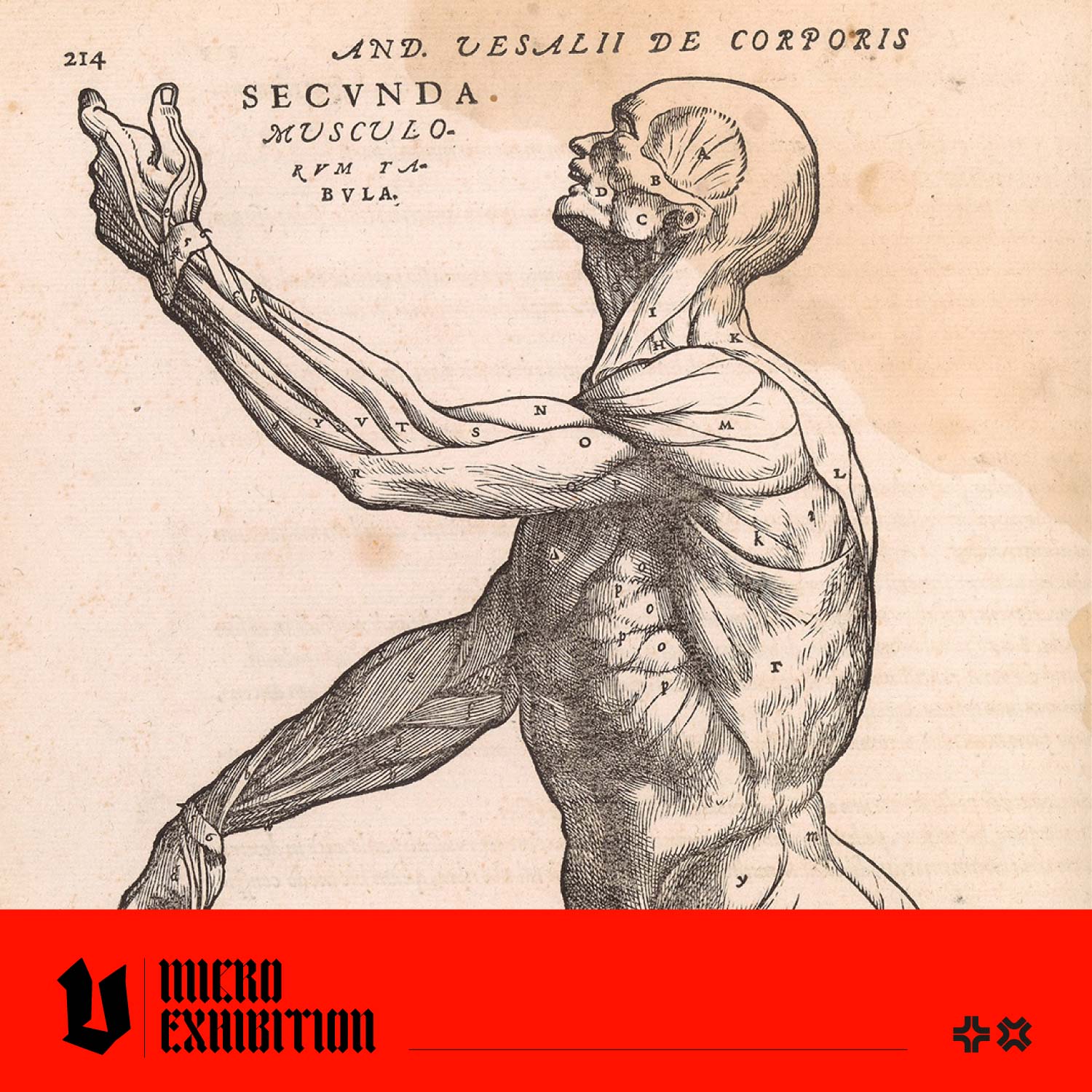
In the 16th century, Andreas Vesalius, a Flemish anatomist, revolutionised the field of anatomical illustration with his groundbreaking work, "De Humani Corporis Fabrica" (On the Fabric of the Human Body). Vesalius's anatomical drawings were characterised by their unprecedented accuracy, intricate detail, and artistic flair. Through meticulous dissections and observations, Vesalius produced illustrations that set new standards for anatomical accuracy and scientific illustration. (For more info, read our article about Andreas Vesalius here!)
Image from De humani corporis fabrica (On the Structure of the Human Body) by Andreas Vesalius (1514–1564)
The Enlightenment and Beyond
The Age of Enlightenment was an intellectual and philosophical movement in Europe in the 17th and 18th centuries. During this period, anatomical illustrations experienced further evolution. Notable anatomists and authors of this period, such as William Cheselden and Bernhard Siegfried Albinus, contributed to the evolution of anatomical studies. With advancements in printing technology, anatomical atlases became more accessible, allowing medical knowledge to spread rapidly. 
Image from Morbid Anatomy, An Image Archive for Artists and Designers
The 19th Century: Rise of Gray's Anatomy
In the 19th century, Dr Henry Gray's 'Gray's Anatomy' emerged as a landmark publication of anatomical illustration. Its engravings, by Dr Henry Vandyke Carter, combine scientific accuracy with artistic elegance and have two very special points of note. Dr Carter dissected the subjects in his illustrations himself, giving him a deeper understanding of their composition, whereas other illustrators would have observed the dissection.
In Britain, the bodies of poor people who died in workhouses or hospitals and couldn't afford a funeral could be used for medical dissection. Dr Carter was a religious man, and his beliefs influenced his illustrations; as historian Ruth Richardson says, "Carter, because of his religious background, sees the human body as the image of God. If you think the human body is the image of God, then you're not going to treat it badly in illustrations; you're going to treat it with dignity. So he looks at these people, who are the poorest of the poor, as though he's mapping the human body as an image of God. If you look at the faces and the postures of the bodies in that first edition you'll see that they've got a dignity to them, which is quite unlike illustrations in contemporary anatomy books."
(Read the original article about Dr. Carter here)
An illustration from the 1918 edition of Gray's Anatomy
Gray's Anatomy, known as 'the doctor's bible' became the standard reference for medical students and professionals, cementing its place in medical literature. It was first published in 1858 and has been revised and republished many times; the current edition (October 2020) is the 42nd edition. 
Image from Morbid Anatomy, An Image Archive for Artists and Designers
The evolution of anatomical illustrations from Vesalius to Gray's Anatomy represents a journey of anatomical discovery made possible by constant innovation, creativity, and scientific progress. While the methods of illustration have evolved over the centuries, the importance of visual representation in understanding the complexities of the human body remains unchanged.
Feeling Inspired?
If you're inspired to delve into the world of anatomical illustration, check out Morbid Anatomy, our latest pictorial archive. This meticulously curated collection of 144 images showcases a macabre gallery of the most visceral anatomical art ever produced, dating from the 16th to the 18th centuries. Designed to inspire and horrify, this book features detailed illustrations of Ecorché figures, brain dissections, flayed figures, and an array of skulls, skeletons, nervous systems, arteries, internal organs and more.


