
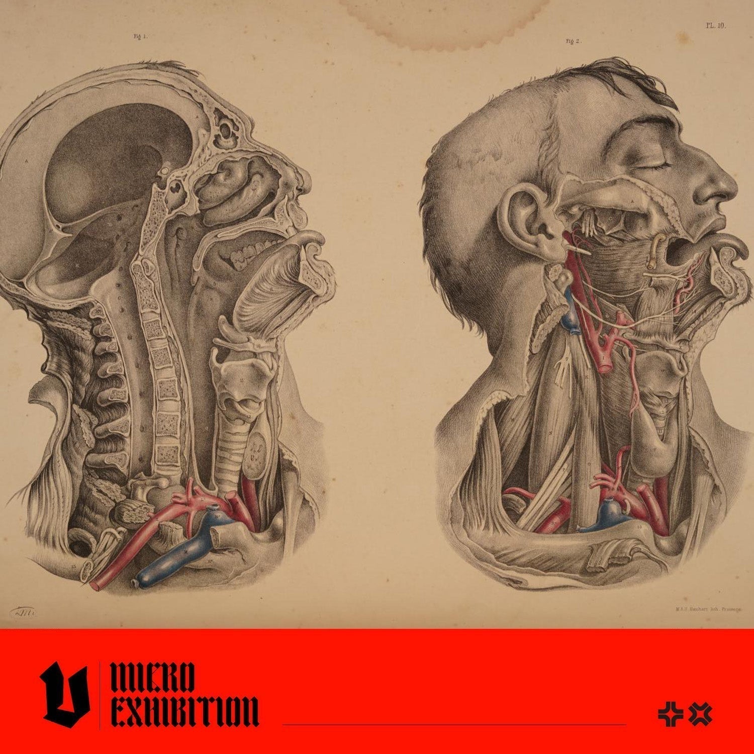
In the mid-19th century, most anatomical atlases depicted the human body like an instruction manual would describe an engine; body parts labelled, components isolated, and all humanity stripped away. Joseph Maclise, a surgeon and illustrator, published an extraordinary book that elevated medical illustration. Today we're learning about his book, 'Surgical Anatomy', and studying some truly mesmerising studies of the human body as you've never seen them before. Let's go!

From Joseph Maclise's Surgical Anatomy, published in London by John Churchill, 1851
Who Was Joseph Maclise?
Joseph Maclise was born in Cork, Ireland, in 1815, the second of seven children. He trained at University College London under the surgeons Robert Liston and Samuel Cooper, and continued his studies in Paris at the École Pratique and the Hôpital de la Pitié. He went on to become a Fellow of the Royal College of Surgeons and established a busy medical practice in London.
He was also the younger brother of Daniel Maclise, a popular history painter known for his monumental study for the Royal Gallery murals at the Palace of Westminster, including The Death of Nelson. The two brothers were close, sometimes living together and travelling to Paris, Lyon, and Naples.

From Joseph Maclise's Surgical Anatomy, published in London by John Churchill, 1851
What Made 'Surgical Anatomy' Different?
Most anatomical atlases of the 18th and early 19th centuries depicted the body clinically, as if it were an isolated organ or a lone limb, but Maclise took a fundamentally different approach. Maclise believed that a well-executed illustration could communicate more directly and precisely than any written description, and his plates prove the point.
His subjects are not cadavers; they are people, young, lifelike, with detailed facial expressions, muscle tone, hair, and elegant poses. They tilt their heads, stretch their arms overhead, lean back as if resting. This was not just artistic flair; it also served a vital surgical purpose. Surgeons needed to understand how muscles, arteries, and nerves shifted in real physical positions during an operation, and they needed more than an image of an isolated organ or a section of the body frozen in a clinical pose to help them prepare for an encounter in the operating theatre.

From Joseph Maclise's Surgical Anatomy, published in London by John Churchill, 1851
Producing the Plates for 'Surgical Anatomy'
Surgical Anatomy was printed in folio format and was large enough to examine anatomical relationships at a near-life-size scale. Maclise personally executed the drawings on lithographic stone, ensuring that no detail was lost in the transition from the dissecting table to the printing press. The first London edition of 1851 contained 35 plates; the second edition of 1856 expanded to 52; and the American edition of 1859 grew to 68.
The plates focused on practical surgical concerns, the ligation of major arteries, hernia repair sites, and deep surgical topographies, and all were rendered with a dramatic beauty that would not have looked out of place on a gallery wall. The style sits somewhere between Romanticism and precise topographical anatomy, and the combination is captivating.
Two of the British edition's plates featured a Black man as an anatomical subject, depicted with the same elegance and humanity as every other figure in the book, and described by the Guardian as potentially the only anatomical drawing of a Black body from the Victorian era. When the American edition was published in Philadelphia in 1859, those plates were altered. As art historian Naomi Slipp notes in British Art Studies, the American printing replaced the figure with a mirror image of his white companion, a change that reflected the racial politics of a country on the cusp of civil war, where slavery was still being practised in the South.

From Joseph Maclise's Surgical Anatomy, published in London by John Churchill, 1851
Why 'Surgical Anatomy' Was Ahead of Its Time
Published just as anaesthesia was beginning to transform operating theatres, 'Surgical Anatomy' represents a high-water mark of pre-digital surgical education. In an era when operations had to be performed with extraordinary speed to minimise patient trauma, a surgeon's mental map of blood vessels and tissue had to be absolute. Maclise's plates provided an invaluable resource, rendered with unforgettable dramatic beauty.
His work was fundamental to the training of surgeons for at least three decades and served as a bridge between two continents. Today, original copies of 'Surgical Anatomy' are prized by rare book collectors, medical historians, and art lovers alike, and beautifully document the intersection of art and medicine.

From Joseph Maclise's Surgical Anatomy, published in London by John Churchill, 1851